History of Present Illness:
A patient in their mid-40’s with no known PMH presents to the hospital with 2 days of left flank pain and urinary frequency. He denies hematuria, fever, vomiting or any other complaints.
Vital Signs & Physical Exam:
Vital signs are normal. Physical exam is also normal except for left CVA tenderness
Initial Diagnostic Testing:
- CBC and CRP: normal
- Chem-7 and UA: normal
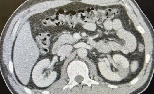
- Imaging: Renal US shows a 2cm L renal cyst, CT contrast shown below

What is the most likely diagnosis?
- A) Renal cyst
- B) Renal stone
- C) Renal infarct
- D) Renal abscess
SCROLL DOWN FOR ANSWERS & 1-MINUTE CONSULT
<<<<<<<<<<<<<<<<<<<<< ADVERTISEMENT & SPACER >>>>>>>>>>>>>>>>>>>>>
THE EMERGENCY MEDICINE POCKETBOOK TRIFECTA

Emergency Medicine 1-Minute Consult, 5th edition
A-to-Z EM Pharmacopoeia & Antibiotic Guide, NEW 5th edition (currently printable pdf only)
8-in-1 Emergency Department Quick Reference, 5th edition
******************************************************************************
<<<<<<<<<<<<<<<<<<<<<<<<< END SPACER >>>>>>>>>>>>>>>>>>>>>>>>>
QUIZ ANSWER:
- A) Renal cyst
- B) Renal stone
- C) Renal infarct – CORRECT – it is the darker area of the kidney that is not contrast enhanced
- D) Renal abscess
1-Minute Consult on this topic: Click HERE and scroll to page 338.
CASE CONCLUSION: Admitted, LDH & D-dimer normal, UA normal except for small protein. Echo showed a dilated RV on preliminary report. Bubble study was not done
CASE LESSONS: When imaging shows a finding that should not be painful (L renal cyst in this case) it is likely a red herring and so you should continue to look for the actual cause if indicated. Although LDH and D-dimer often elevated with renal infarct, this is not always the case.
***
