History:
A patient in their early-60’s with a history of HTN, high cholesterol and chronic back pain is brought to the hospital by ambulance for sudden onset of back and left flank pain radiating to both legs. He denies any injury but does do a lot of lifting at work. He states that he also has a history of kidneys stones but states “this is different and it feels like my sciatica and kidney stones at the same time, but it’s both legs this time.” He is requesting Dilaudid IV. He denies fever, trouble urinating, leg swelling, or other complaints.
Exam:
Pulse is in the low 60’s, BP is 138/77 and other vitals are normal. Patient is slightly diaphoretic and can wiggle his toes but not lift his legs or knees off the gurney. Pulses feel strong and symmetric in both arms but diminished though symmetric in both legs with palpable femoral pulses but not the DP pulses bilaterally.
Labs: creatinine elevated at 1.7, which was new, microscopic hematuria. Other labs were normal or at patient’s baseline.
POCUS done on arrival: No hydronephrosis. The abdominal aorta was gassed out. A view of the right femoral artery is shown below.
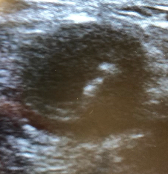
To see the video, click here
What is the most likely diagnosis?
- A) Cauda equina
- B) AAA
- C) Aortic dissection
- D) Kidney stone
“BRADY” DOWN FOR THE EKG ANALYSIS & 1-MINUTE CONSULT
<<<<<<<<<<<<<<<<<<<< ADVERTISEMENT & SPACER >>>>>>>>>>>>>>>>>>>>
******************************************************************************
******************************************************************************
THE EMERGENCY MEDICINE POCKETBOOK TRIFECTA

Get one of our publications, all designed specifically for Emergency Care Providers:
Emergency Medicine 1-Minute Consult, 5th edition
A-to-Z EM Pharmacopoeia & Antibiotic Guide, 5th edition
8-in-1 Emergency Department Quick Reference, 5th edition
*****************************************************************************
<<<<<<<<<<<<<<<<<<<<<<<<< END SPACER >>>>>>>>>>>>>>>>>>>>>>>>>
QUIZ ANSWER: What is the most likely diagnosis?
- A) Cauda equina – can affect both legs, but shouldn’t affect pulses
- B) AAA – would usually affect arms and legs equally through hypotension
- C) Aortic dissection – CORRECT. Image shows dissection in the femoral artery
- D) Kidney stone – POCUS still misses about 20%, but stones shouldn’t affect pulses, though they could be chronic
Case Outcome: CT showed a type B aortic dissection from just below right subclavian into right femoral artery causing infarct of entire left kidney and extending into right leg. Eventually left leg got pulse back. Unclear if flap had occluded it or there was an embolism that dissolved. Went to IR. CT, consultation and treatment all occurred very rapidly due to bedside ultrasound confirming the diagnosis within minutes of arrival.
Case Lessons:
- Always check DP pulses in patients with leg symptoms
- Dissection doesn’t always have chest pain
- Bedside ultrasound helps expedite the diagnosis in critical cases. Most dissections go into at least one leg so in addition to looking for a pericardial effusion or an arch dissection consider looking at the abdominal aorta and/or femoral artery. The femoral artery may be the easiest to see and therefore may be the most sensitive place to make the diagnosis.
1-Minute Consult: Click HERE and scroll to page 18