History of Present Illness:
A man in his mid 20’s with a history of type 1 diabetes and JRA presents to the ED for 1-2 weeks of gradually worsening finger deformity after an injury at football practice. He said he hyperextended the whole finger. It’s not really painful but is a bit swollen and is gradually getting stiffer.
Vital Signs & Physical Exam:
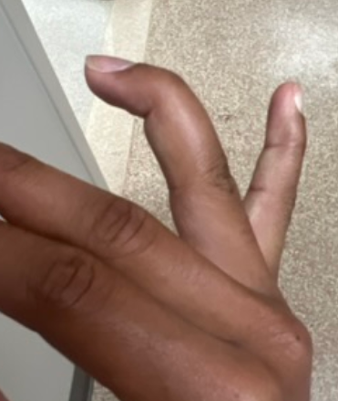
See finger below. Flexion was limited to about 20% of normal at the PIP joint. He could not extend the DIP joint
Initial Diagnostic Testing:
- Imaging: x-ray of finger was normal
What does the image show?
- A) Mallet finger
- B) Boutonniere deformity
- C) Swan neck deformity
- D) Jersey finger
SCROLL DOWN FOR ANSWERS & 1-MINUTE CONSULT
<<<<<<<<<<<<<<<<<<<< ADVERTISEMENT & SPACER >>>>>>>>>>>>>>>>>>>>>
THE EMERGENCY MEDICINE POCKETBOOK TRIFECTA

Emergency Medicine 1-Minute Consult, 5th edition
A-to-Z EM Pharmacopoeia & Antibiotic Guide, NEW 5th edition
8-in-1 Emergency Department Quick Reference, 5th edition
******************************************************************************
<<<<<<<<<<<<<<<<<<<<<<<<< END SPACER >>>>>>>>>>>>>>>>>>>>>>>>>
What does the image show?
- A) Mallet finger – Very Close – looks similar but this is a bit hyperextended at the PIP joint and his PIP flexion was limited
- B) Boutonniere deformity – would be forced flexion at PIP and extension at DIP
- C) Swan neck deformity – CORRECT
- D) Jersey finger – inability to fully flex DIP joint
CASE CONCLUSION: splinted in extension and had f/u arranged with ortho hand