
Triage: Has cramping in lower abd radiating to sides and upper quadrants. 10/10 pain with vomiting. No diarrhea or blood.
History of Present Illness: female in her late 70’s with past medical history of osteoporosis presents with several days of constipation and 1 day of severe generalized abdominal pain in addition to 4 bouts of nonbilious non-bloody emesis. She states she had a bowel movement that was small and firm and non-bloody this morning but since then has not been passing flatus and states that the pain radiates up towards her epigastric region. She denies fevers or chills or other symptoms
Vital Signs: T: 36.5 °C (Oral) HR: 60 (Monitored) RR: 18 BP: 165/82 SpO2: 98%
Physical Exam: Uncomfortable appearing and retching generalized abdominal distention and tenderness with pain out of proportion to exam
Initial Diagnostic Testing:
- Labs: abdominal labs with CBC, PMN%, CMP and lipase in addition to urinalysis were ultimately within normal limits. Lactate obtained and was initially elevated at 2.8 and after 1 L of NS it down-trended and normalized to 1.8. WBC on repeat doubled from 6 to 13 and PMN% went from 60% to 90%
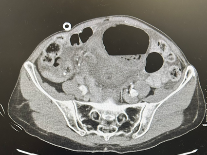
- Imaging: CT cut shown below
SCROLL DOWN FOR ANSWERS & 1-MINUTE CONSULT
What is the most likely diagnosis?
- A) Perforated viscus
- B) Mesenteric Ischemia
- C) SBO
- D) LBO
<<<<<<<<<<<<<<<<<<<<< ADVERTISEMENT & SPACER >>>>>>>>>>>>>>>>>>>>>
THE EMERGENCY MEDICINE POCKETBOOK TRIFECTA

- Emergency Medicine 1-Minute Consult, 5th edition
- A-to-Z EM Pharmacopoeia & Antibiotic Guide, NEW 5th edition
- 8-in-1 Emergency Department Quick Reference, 5th edition
******************************************************************************
<<<<<<<<<<<<<<<<<<<<<<<<< END SPACER >>>>>>>>>>>>>>>>>>>>>>>>>
What is the most likely diagnosis?
- A) Perforated viscus
- B) Mesenteric Ischemia
- C) SBO
- D) LBO – Correct: Had a Cecal Volvulus that was missed on this CT but diagnosed on a subsequent CT
CT read: IMPRESSION:
1. Moderate fluid in the abdomen. Source not identified.
2. Unopacified tissue in the right pelvic sidewall, unclear if representing unopacified bowel or other such as adenopathy or mass; significance not clear.
3. No evidence of bowel obstruction.
4. Small hiatal hernia.
5. Moderate stool in the colon.
1-Minute Consult on this topic: Click HERE and scroll to proper page
CASE CONCLUSION: CT next day showed increase in fluid and a cecal volvulus. She was taken to the OR with no e/o perforation but did require a right hemicolectomy.
CASE LESSONS:
- Volvulus: Closed loop obstruction so risk for necrotic bowel. XR: 30%, UGI: 95%
- Gastric: Risks: diaphragmatic defect Rx: IV fluid, consult GI & surgery
- Sigmoid: Risks: age, neuro-psych dz, constipation, H1 Rx: IVF, detorse by GI >OR
- Cecal: Risks: congenital (typically a young adult) Rx: IV fluids, OR